Dense Breast Imaging: Latest Techniques and Tools
Breast density is more than just a radiological observation; it is a critical factor in women's health and early cancer detection. With the FDA’s recent requirement for uniform breast density reporting, the conversation around screening protocols for dense tissue has never been more relevant.
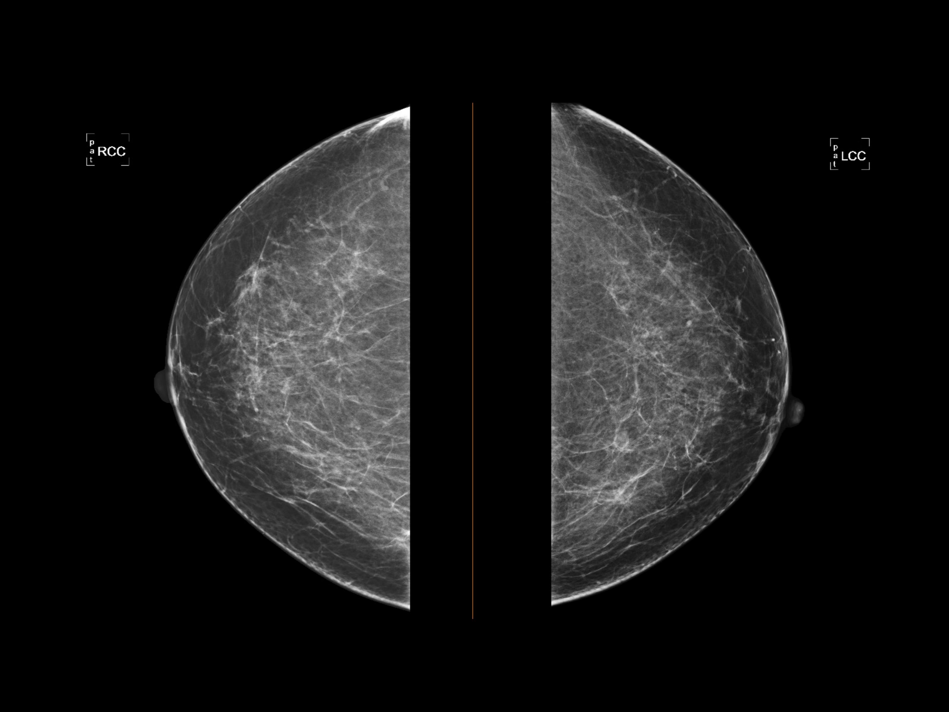
For imaging professionals, understanding the nuances of dense breast tissue and the evolving technology used to assess it is essential. Dense tissue, composed of more glandular and fibrous connective tissue than fatty tissue, poses two significant challenges. First, it is an independent risk factor for developing breast cancer. Second, it creates a masking effect on traditional mammograms. Since both dense tissue and tumors appear white on X-rays, finding a lesion can be akin to looking for a snowball in a blizzard.
Fortunately, the landscape of breast imaging is advancing rapidly. From 3D tomosynthesis to molecular imaging, new tools are improving sensitivity and specificity, helping clinicians catch cancers that might otherwise remain hidden.
The Limitations of 2D Mammography
Digital mammography remains the gold standard for screening, but its limitations in dense breasts are well-documented. Sensitivity can drop significantly when screening women with heterogeneously dense (BI-RADS C) or extremely dense (BI-RADS D) tissue.
While 2D mammography is efficient and effective for fatty breasts, the superposition of tissues in a 2D image often obscures small masses. This limitation has driven the rapid adoption of supplemental screening methods and advanced imaging modalities designed specifically to look through the density.
Digital Breast Tomosynthesis (DBT)
Digital Breast Tomosynthesis, commonly known as 3D mammography, has become the new standard of care in many facilities. Unlike a standard 2D mammogram, which captures a single flat image, DBT takes multiple low-dose X-ray exposures from different angles. Computer software then reconstructs these images into a series of thin slices.
This "slicing" capability allows radiologists to scroll through the layers of breast tissue, significantly reducing the masking effect of overlapping tissue. Studies have consistently shown that DBT increases cancer detection rates in invasive cancers and reduces unnecessary recall rates, offering a clearer picture for patients with dense tissue.
Whole Breast Ultrasound
Ultrasound has long been the primary problem-solving tool for palpable lumps or focal asymmetries. However, it has gained traction as a supplemental screening tool for asymptomatic women with dense breasts.
Automated Breast Ultrasound (ABUS) systems have standardized this process. Unlike handheld ultrasound, which is operator-dependent and time-consuming, ABUS uses a wide-field transducer to scan the entire breast automatically. This provides a consistent, reproducible 3D volume of the breast tissue, allowing radiologists to detect small, invasive cancers that are mammographically occult, i.e., a breast cancer is present but not visible on a mammogram.
Breast MRI
For women at high risk—typically defined as a lifetime risk of 20% or more—Breast MRI remains the most sensitive supplemental screening tool available. It is not hindered by tissue density and utilizes contrast dye to highlight angiogenesis (increased blood flow) associated with tumors.
While highly effective, MRI is expensive and requires intravenous contrast (a fluid injected into a vein to make blood vessels, organs, and abnormalities more apparent), making it less suitable for the general screening population. However, abbreviated MRI protocols—often called "Fast MRI"—are currently being studied as a way to reduce scan times and costs while maintaining high diagnostic accuracy for intermediate-risk women with dense breasts.
Emerging Technologies: MBI and CEM
Two other modalities are making significant headwinds in the fight against missed detections in dense tissue:
Molecular Breast Imaging (MBI)
MBI is a nuclear medicine technique that requires the injection of a radioactive tracer. Because cancer cells differ metabolically from normal cells, they absorb more of the tracer. The MBI camera detects this activity, revealing tumors even in extremely dense breasts. It is a powerful functional imaging tool, though radiation dose considerations remain a topic of ongoing optimization.
Contrast-Enhanced Mammography (CEM)
CEM combines the accessibility of digital mammography with the functional insights of MRI. By using an iodinated contrast agent typically used in CT scans, CEM highlights areas of increased blood flow. It is faster and more affordable than MRI and can be performed on modified digital mammography units, making it an attractive option for imaging centers looking to expand their diagnostic capabilities without installing a magnet.

Advance Your Career with AMN Healthcare
Staying current with these technological advancements is vital for providing the best patient care, but it also opens doors for your career. As imaging technology continues to evolve, professionals who embrace these tools both enhance diagnostic accuracy and position themselves as leaders in the field. Partnering with organizations like AMN Healthcare ensures access to mentorship, transparent pay, and exclusive opportunities that support your growth. By mastering the latest breast imaging techniques, you can make a meaningful impact on patient outcomes while advancing your professional journey.